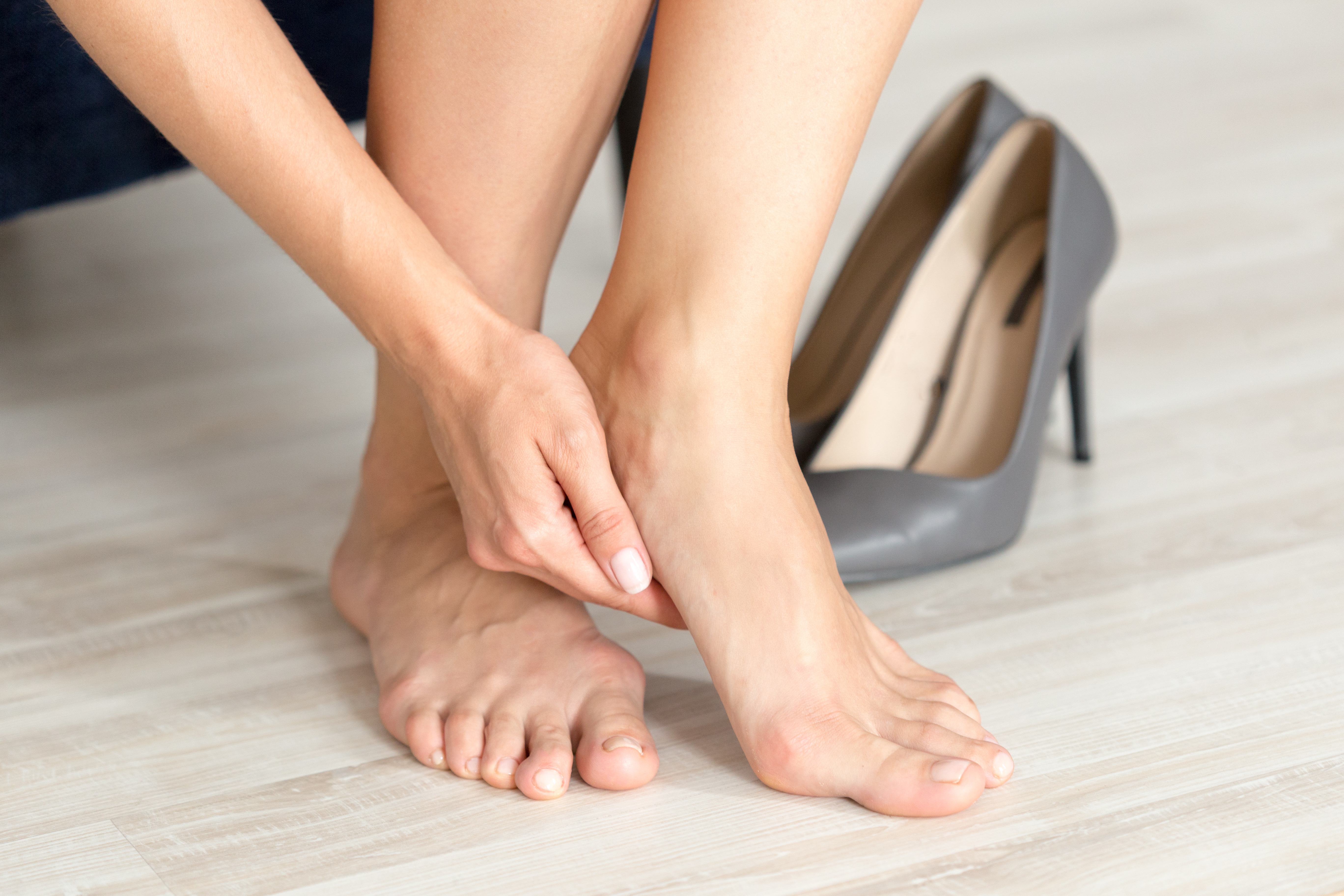
With heel pain accounting for 15% of all foot-related consultations, keeping informed about best practices is vital.
This overview summarises recent evidence-based guidelines and expert consensus on heel pain management, offering practical tips for tailoring interventions. Whether you are a physiotherapist, podiatrist, general practitioner or lower limb specialist, this blog aims to help you navigate current evidence while considering the unique circumstances of each patient.
Summary
Heel pain is common and is most often due to plantar fasciitis.
A variety of evidence-based conservative treatments are beneficial in improving heel pain due to plantar fasciitis. These include key strategies such as stretching, strengthening exercises, taping and education and can also include footwear modifications, orthotic support, corticosteroid injections, shockwave therapy, dry needling, night splints, low-light laser therapy and electrotherapy.
Treatment should be individualised according to each patient’s presentation, lifestyle and needs. Cost and accessibility are also important considerations.
Understanding plantar heel pain
Plantar heel pain is one of the most common complaints seen in primary care and musculoskeletal clinics worldwide. It is most frequently caused by plantar fasciitis, a condition characterised by degeneration of the plantar fascia, which typically causes pain under the heel, especially with a person’s first steps after rest or prolonged sitting. It most commonly affects people aged 40–60 years, particularly runners and those in occupations that involve prolonged standing.
It is important to be aware of other heel pain causes, which include heel fat pad syndrome, heel spur syndrome, nerve irritation, calcaneal stress fracture, inflammation of the Achilles tendon and bone or soft tissue tumours.
Best practices for diagnosis and management of plantar heel pain
Clinical guidelines can help clinicians diagnose and manage heel pain. Consensus statements can provide additional expert insights into practical matters or areas of uncertainty.
This summary of key publications is intended to help you incorporate evidence into practice. Extra detail on diagnosis and conservative management follows.
Recent guidance and expert consensus
American Physical Therapy Association (APTA) and Journal of Orthopaedic & Sports Physical Therapy (JOSPT) Clinical Practice Guidelines, 2023: Provides evidence-based recommendations for diagnosis and treatment of plantar fasciitis.
Treatment should include manual therapy, plantar fascia-specific stretching, calf stretching, strengthening, and neuromuscular re-education, dry needling, foot taping (in conjunction with other physical therapy treatments for short-term improvements), and night splints (for patients who consistently experience pain with the first step in the morning).
Optional adjuncts include orthoses, electrotherapy, low-light laser therapy, phonophoresis and patient education.
Plantar heel pain: A best practice guide, 2021: Published in the British Journal of Sports Medicine, this guide to management of plantar heel pain was informed by a systematic review, expert clinical reasoning and patient values.
A core approach should combine stretching, taping and individualised education. These should be used together for approximately 4–6 weeks before adjunctive interventions such as extracorporeal shock wave therapy (ESWT) or orthoses are considered.
National Institute for Health and Clinical Excellence (NICE) Clinical Knowledge Summary, 2024: Provides an evidence-based stepwise primary care pathway for plantar fasciitis (content available in the UK only).
American College of Foot and Ankle Surgeons (ACFAS) Clinical Consensus Statement, 2017: Provides consensus statements based on a review of evidence and the experience of an expert panel.
The panel agreed that, in most cases, plantar fasciitis is diagnosed using history and physical examination findings. Routine use of radiographs is unnecessary for the diagnosis of nontraumatic plantar fasciitis.
The panel agreed that the following are all safe and effective in the treatment of plantar fasciitis: biomechanical support (including taping or strapping, over-the-counter insoles, custom foot orthoses and weight management counselling), stretching, corticosteroid injections (CSIs) and ESWT.
ACFAS suggests that, rather than following a treatment hierarchy, treatment should be customised to each patient’s symptoms (including chronicity and severity), lifestyle and level of activity. Treatment should consider a patient’s specific expectations and physical requirements. Treatment must also be cost-conscious and should consider accessibility within the local healthcare system.

Practical assessment strategies
History taking
A thorough patient history is essential for diagnosing plantar heel pain and distinguishing it from other causes of foot discomfort. Key elements include:
Pain characteristics
Risk factors
Previous treatments
Red flags for referral or further investigation
Physical examination
The physical exam helps to confirm the diagnosis, can identify potential biomechanical contributors and can be used to assess the impairment of body function.
Key components:
Palpation
Functional tests (eg windlass test and tarsal tunnel tests)
Range of motion (eg ankle dorsiflexion)
Gait and foot posture
Imaging (if indicated)
Radiographs can help rule out other causes of pain and should be ordered in cases of trauma, pain out of the ordinary or recalcitrant pain that is not responding to appropriate conservative treatment.
Conservative treatment options
This section provides an overview of some commonly recommended interventions.
Patient education: Education includes advice on load management, activity modification and supportive footwear.
Stretching and exercise therapy: Daily plantar fascia-specific and calf stretches reduce pain and speed recovery. As symptoms improve, progressive strengthening of the foot and calf muscles helps address biomechanical deficits.
Taping: Foot taping techniques can provide short-term relief by reducing plantar fascia strain.
“Taping can be used to support the arch and rest the plantar fascia in the short term. It can also be used as a test to determine whether the patient would do well with a more controlling insole or custom orthotic.” (ACFAS, 2023)
Footwear modification: Supportive shoes that allow a small rearfoot to forefoot drop to reduce compression should be encouraged.
Orthotic devices: Prefabricated, custom or mouldable orthoses can redistribute load and improve biomechanics. Orthotics can provide significant pain relief for patients with plantar heel pain, notably in the moderate term (7–12 weeks), but they are best considered alongside other adjuncts.
APTA/JOSPT recommend considering orthotics in combination with other treatments, including,
“use of over-the-counter/prefabricated or custom foot orthoses that support the medial arch and/or provide cushion to the heel region, especially in individuals with Foot Posture Index-6 scores indicating excessive pronation and/or who positively respond to antipronation taping.” (APTA/JOSPT 2023)
Night splints: Particularly helpful for patients with morning pain or stiffness, as they maintain the plantar fascia in a lengthened position overnight.
ESWT: ESWT may stimulate healing and has been shown to reduce pain.
Injections: CSIs may provide short-term relief but carry risks, including plantar fascia rupture. Alternatives such as platelet-rich plasma are being explored but require more evidence.
Exercise therapy beyond stretching: Resistance training for the musculature of the foot and ankle can be beneficial as part of a multimodal treatment strategy.
Surgical intervention
Surgery should be reserved for chronic, refractory cases for which appropriate conservative treatment for ≥6 months has failed.
Managing complex cases
Some patients have persistent (>3–6 months) or atypical plantar heel pain due to multifactorial risk factors such as obesity, systemic inflammatory disease or occupational demands. For these individuals, a multidisciplinary approach involving physiotherapy, podiatry and orthopaedics may be necessary. Care plans should address risk factors and should be reviewed regularly to help optimise outcomes and prevent chronicity.
Optimising outcomes
Set expectations: Recovery often takes several months, but most patients experience symptomatic relief with 3–6 months of conservative treatment.
Encourage adherence: Daily stretches and consistent use of conservative interventions are key.
Review regularly: Reassess foot biomechanics, adherence and symptom progression.
Combine strategies: Multimodal care is generally most effective.
Conclusion
Numerous evidence-based publications provide support to clinicians in the diagnosis and management of plantar heel pain. For management of plantar heel pain, education, stretching, strengthening, taping and footwear modification are recommended as key components of care. Other conservative interventions, including night splints, orthotic devices and ESWT, are supported by evidence and can be included in individualised care plans. For individualised care, plantar heel pain management should be grounded in guidelines but tailored to each patient’s biomechanics, lifestyle, expectations and access to healthcare.
How could Formthotics help your patients with plantar heel pain?
Clinical studies have shown that Formthotics orthotics provide heel pressure relief in patients with plantar heel pain.
Improvements in pain have been observed, particularly in the mid-term (~12 weeks).
For example, when Formthotics were compared with CSIs, pain improvements were better with CSI at week 4 but better with Formthotics by week 12.*
The talonavicular support built into the Formthotics’ unique contour is designed to act in a similar way to low-dye taping to reduce the negative effects of excessive pronation on the plantar fascia.
In cases where this support increases compressive forces at the medial calcaneal tubercle, a clinician may choose one of several options to mitigate this: grind away part of the teardrop shape beneath the orthotic, cut a fascial groove in the top surface of the orthotic or add a medial rearfoot post, using one of the Formthotics additions, to shift forces away from the medial aspect of the foot.
Formthotics’ arch support and deep heel cup reduce pressure on the calcaneal tuberosity by increasing the calcaneal pitch angle and by preventing the escape of the plantar fat pad, respectively.
Our variety of products mean that support and cushioning can be individualised.
The Dual Density orthotics range provides a firm layer of support (of various densities) with a top layer for cushioning.
The ShockStop model uses a unique shock-absorbing foam that can help reduce compressive forces on the plantar fascia at the attachment point of the medial calcaneal tubercle.
The Single Density orthotics range can be useful if the clinician needs to customise treatment by the addition of soft pads or a specific top layer.
The Formthotics Medical System includes a range of additions which can be easily added for customisation and optimisation of treatment by the clinician.
Formthotics are heat-moulded in the clinic within minutes and can be a one-off, affordable, non-invasive treatment to provide immediate comfort and support, allowing the patient to remain active and maintain access to all the well-known physical, mental, and psychosocial benefits of exercise.
This content is for general information only and does not replace the advice of a qualified healthcare professional, localised best practice and standards of care. Information in this material is not a substitute for clinical assessment and relevant information pertaining to specific interventions. For personal advice, consult a healthcare professional. Availability of treatments and their indications may vary by country. For the best outcomes and full support, use Formthotics only with approved accessories as listed in the Declaration of Conformity.
*Whittaker GA, et al. 2019: In 103 patients with plantar heel pain, improvements in pain versus baseline were observed with both Formthotics and CSI, and these were greater with Formthotics (the difference was statistically significant but did not meet criteria for clinically meaningful difference).
References
Babatunde OO, et al. Comparative effectiveness of treatment options for plantar heel pain: a systematic review with network meta-analysis. Br J Sports Med. 2019;53:182–194.
Bonanno DR, et al. Pressure-relieving properties of various shoe inserts in older people with plantar heel pain. Gait Posture. 2011;33(3):385–389.
Caratun R, et al. Stubborn heel pain. Treatment of plantar fasciitis using high-load strength training. Can Fam Phys. 2018;64:44–46.
Chia KK, et al. Comparative trial of the foot pressure patterns between corrective orthotics, Formthotics, bone spur pads and flat insoles in patients with chronic plantar fasciitis. Ann Acad Med. 2009;38(10):869–875.
Del Duchetto F, et al. Can foot orthoses benefit symptomatic runners? Mechanistic and clinical insights through a scoping review. Sports Med Open. 2024;10(1):108.
Koc TA Jr, et al. Heel pain - plantar fasciitis: Revision 2023. J Orthop Sports Phys Ther. 2023;53(12):CPG1–CPG39.
Landorf KB, et al. Effectiveness of foot orthoses to treat plantar fasciitis. Arch Intern Med. 2006;166(12):1305–1310.
Latt LD, et al. Evaluation and treatment of chronic plantar fasciitis. Foot Ankle Orthop. 2020;5(1):2473011419896763.
Morrissey D, et al. Management of plantar heel pain: a best practice guide. Br J Sports Med. 2021;55:1106–1118.
National Institute for Health and Care Excellence. Clinical Knowledge Summary: Plantar Fasciitis. London: NICE; 2024.
Ohuchi H, et al. Changes in calcaneal pitch and heel fat pad thickness in static weight bearing radiographs while wearing shoes with arch support and heel cup orthotics. Asia Pac J Sports Med Arthrosc Rehabil Technol. 2019;17:21–24.
Riddle DL, et al. Risk factors for plantar fasciitis: a matched case-control study. J Bone Joint Surg Am. 2003;85(5):872-877.
Rio E, et al. Heel pain: a practical approach. Aust Fam Physician. 2015;44(3):96–101.
Schneider HP, et al. American College of Foot and Ankle Surgeons Clinical Consensus Statement: Diagnosis and treatment of adult acquired infracalcaneal heel pain. J Foot Ankle Surg. 2018;57(2):370–381.
Trojian T, et al. American Academy of Family Physicians. Plantar fasciitis. Am Fam Physician. 2019;99(12):744–750.
Whittaker GA, et al. Effectiveness of foot orthoses versus corticosteroid injection for plantar heel pain: The SOOTHE randomized clinical trial. J Orthop Sports Phys Ther. 2019;49(7):491–501.
Whittaker GA, et al. Foot orthoses for plantar heel pain: a systematic review and metaanalysis. Br J Sports Med. 2018;52(5):322–328.
Related Articles
:format(webp))
Heel Pain and Orthotic Treatment
Understand the causes of heel pain and how orthotic therapy supports effective relief and recovery.
:format(webp))
Plantar Fasciopathy
Heel pain affecting performance? See how effective orthotic care turned things around.
:format(webp))
Formthotics treat Plantar Fasciitis: RCT Results
Effectiveness of foot orthoses to treat plantar fasciitis: a randomized trial